
Dental X-rays are a way of answering questions you cannot answer by looking. A tooth can appear perfectly healthy from the outside while decay spreads between it and its neighbor, or while the bone that holds it is quietly thinning. An image shows what is happening below the surface, between the teeth and inside the jaw.
The useful question is rarely "Do I need X-rays?" It is "What are we trying to find out, and would the answer change what we do?" Those questions decide which image to take, how many and how often. Someone with a new ache needs a different image than someone with ten years of stable checkups.
This page explains what each type of dental image shows, why there is no one-size schedule, how exposure is kept low, and how to talk with us about imaging so you understand every picture we take.
What X-rays can show that an exam cannot
During an exam we can see the visible surfaces of your teeth and the gums around them. We cannot see:
- Decay between teeth, where two back teeth touch and a toothbrush and even a mirror cannot reach. This is the most common place for a cavity to hide.
- Decay under existing fillings and crowns, which can grow for years before a filling falls out or a tooth starts to hurt.
- Bone level around the roots. Gum disease destroys bone before gums visibly recede; the bone line on an X-ray is how we track it.
- The tips of the roots, where an infection from a dying nerve forms a dark area long before swelling appears.
- Teeth that have not come in yet, including wisdom teeth and a child's developing permanent teeth.
- Cracks, cysts, extra or missing teeth and unusual root shapes that affect how a tooth can be treated.
Finding these things early is the difference between a small filling and a root canal, or between treating gum disease and losing a tooth to it. That is why imaging sits at the center of general and family dentistry.
Different images answer different questions
Not all dental X-rays are the same, and we do not take all of them on everyone.
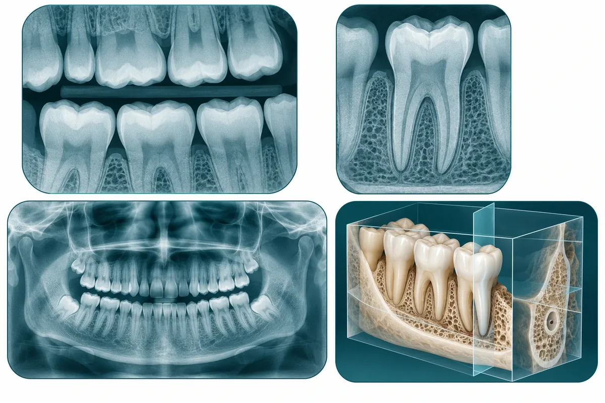
| Image | What it shows | When it is used |
|---|---|---|
| Bitewings | The crowns of the upper and lower back teeth on one side, plus the top of the bone | Checking for decay between teeth and early bone loss; the standard "checkup" image |
| Periapical | One or two whole teeth from crown to root tip and the bone around them | A tooth that hurts, is loose, has had a root canal, or is being planned for a crown or extraction |
| Panoramic | Both jaws, all teeth, the sinuses and jaw joints in one wide view | Wisdom teeth, developing teeth in children, planning extractions, dentures or implants, a broad first look |
| Cone-beam 3D (CBCT) | A three-dimensional view of a region of the jaw, in thin slices | Measuring bone for implants, locating nerves and sinuses, complex root canals and impacted teeth |
Small images give fine detail of a few teeth. The panoramic view trades detail for coverage. 3D imaging answers questions that need depth, such as how much bone is available for an implant, and is not part of a routine exam. Our guides to wisdom teeth removal and dental implant candidacy explain where those broader images fit.
Why there is no single schedule for everyone
You may have heard "X-rays once a year" as if it were a rule. It is not. The right interval depends on how likely something is to change between visits.
Reasons you might need checkup images more often:
- new cavities in the last few years
- a lot of fillings and crowns to monitor
- gum disease that is being treated or watched
- dry mouth from medication, frequent snacking or sugary drinks
- teeth still developing, since children's mouths change quickly
Reasons you might need them less often:
- several years without new decay
- healthy gums with stable bone on previous images
- few or no restorations
- good home care and low-sugar habits
Symptoms override the schedule. If a tooth starts aching or a filling feels different, a targeted image is appropriate even if your routine images were taken last month. If you are visiting for the first time, we may need a baseline set so that future images have something to be compared against. Our exams and cleanings guide explains how imaging fits into the first visit.
Using images you already have
If you have had dental X-rays in the last few years, ask that office to send them to our Cleburne office before your appointment. Most offices can transfer digital images in a day or two. Good recent images can mean fewer new ones.
Whether old images are enough depends on three things: how recently they were taken, whether they cover the area in question, and whether they are sharp enough to read. A blurry bitewing from four years ago cannot tell us what is happening between your molars today. A clear panoramic image from last year may be perfectly good for planning. We will tell you which is which, and why.
Radiation, safety and pregnancy
Dental X-rays use ionizing radiation, so every image should have a reason behind it. The dose from modern digital sensors is small: a set of four bitewings is in the same range as the natural background radiation a person receives in a day or so of ordinary life. Digital sensors need far less exposure than the film many adults remember, and the beam is aimed at a small area.
We keep exposure low by taking images only when they answer a question, by using the smallest image that does the job, and by reusing good recent images rather than repeating them.
If you are pregnant or think you might be, tell us. Dental imaging is considered safe in pregnancy when it is needed, and an untreated infection carries its own risks to you and the baby. Routine images can often wait until after delivery; images needed to diagnose pain or swelling should not. We will talk it through with you rather than making the decision by default.
What an X-ray cannot do on its own
An X-ray is one piece of information, not a verdict. We read it alongside your symptoms, what we see and feel in the mouth, gum measurements and sometimes tests of how a tooth responds to cold or tapping. A dark spot on an image does not automatically mean a tooth is lost, and a normal-looking image does not explain away pain. Some cracks, for example, never show on a two-dimensional X-ray at all.
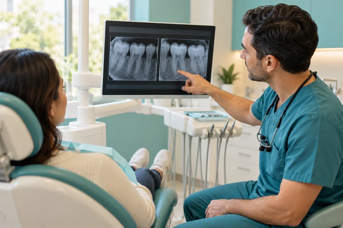
That is why we show you the image and point to what we are seeing. If a recommendation rests on a finding in an X-ray, you should be able to see that finding yourself.
Sources and further reading
General information, not a diagnosis. Read how we create and review our dental guides.